
I don’t know about you, but sometimes I catch myself staring at something and start to wonder how it’s possible I can glean so much information from what is essentially photons bouncing around the room.
A basic description of sight will tell you something like this: photons of light bounce off objects and travel into your eye through the pupil. Your lens directs the incoming photons onto the retina at the back of the eye, where special cells called rods and cones capture them and convert the light energy into nerve signals for your brain to interpret.

Whether these rod and cone cells can detect a photon or not depends on its energy [note 1], and in the grand electromagnetic scheme of things, humans are pretty much blind. Most photons that enter our eyes go completely unnoticed – we can’t detect them unless they have a specific energy, which we call the “visible range”. There is nothing particularly special about this tiny sliver of the electromagnetic spectrum, other than the fact it helpfully (but not coincidentally) corresponds to photon energies most strongly emitted by our sun. If we were to meet aliens who came from a planet with a star like blue Bellatrix or red Antares, they would probably have very different experiences of the universe to us, because their visible range (assuming they had any sort of eyes at all) would be centred in quite a different part of the spectrum.
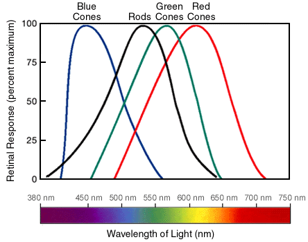
Within the visible range, we have the ability to detect colour. Photons with less energy translate into red and orange, and photons with more energy appear blue and violet. Humans have three types of cone cell which specialise in detecting different, but overlapping photon energies. These cone cells work best in brighter environments. When light levels drop, the rod cells take over. You only have one type of rod, so colour-vision all but disappears, but they have the benefit of being a lot more sensitive. But how can a cell detect individual photons?
Let’s get biochemical
The key protein in rod cells is rhodopsin, which sits inside the cell membrane surrounding the rod cells. Inside it is a smaller molecule called retinal, which is chemically similar to Vitamin A and visually similar to a tiny caterpillar attached by its tail to the inside of the rhodopsin protein… to me anyway.

Normally, the caterpillar is happily bent in the middle, but when a photon with just the right energy hits it, its back-end flips around so it fully stretches out. The head and neck of the caterpillar are actually trapped in place by the protein surrounding it, so it has to move its tail, and with it, some of the protein. The entire rhodopsin protein shifts and contorts, taking on a new shape. A different shape means a different function, and in this case, a different name: metarhodopsin II.
And now we initiate something like the butterfly effect (or in this case, the caterpillar effect), where one small shift brings about big changes within the cell and beyond. One protein molecule has detected a photon of light successfully, and somehow it’s got to spread the word throughout the whole cell. Here’s how it works:

If you want a message to spread to as many people as possible, you have to amplify it, i.e. hire more messengers and send more copies. In cell biology it’s no different. One activated protein can switch on many others and the signal spreads. The final result is that the channels which usually let positively charged ions like calcium (Ca2+) and sodium (Na+) into the cell get blocked. This changes the electric charge between the inside and outside of the cell, and actually starts a nerve impulse destined for the brain.
From eye to brain
The retina at the back of the eye contains many layers of cells stacked on top of each other. These include the rods, cones and many types of nerve cell to carry the signals to the brain.
Given that the rods and the cones’ main job is to detect light, one might make the prudent prediction that these light-detection cells would be found at the front of the retina… y’know, where all the light is.
What we actually find is rod and cone cells stuck right at the back of the stack. So light has to travel past all the clutter of the nerve cells headed for the brain, and the blood vessels supplying them with oxygen. Once the nerve signal gets produced, it has to travel all the way to the front of the retina again, where the nerves all clump in one off-centre spot so they can get to the back of the eye again, and then on to the brain!

If it sounds convoluted, that’s because it is! It’s unclear whether this strange set-up is a tolerable-but-unhelpful evolutionary quirk, or whether it does actually confer some benefit over the “logical” solution. The eye has evolved many times in the animal kingdom, so it’s possible to compare and contrast. We know octopi have eyes which are the “right way around”, and so do not have blind spots.
There are plenty more things where our vision would get a “could try harder” grade, and that’ll be the topic of my next sensory post.
Em x
Bonus links:
1) Click here for a video allowing you to prove to yourself that your eyes are built “backwards” by seeing the blood vessels in front of your rods and cones, as well as the blind spot.
2) Click here for a Vsauce video considering whether your interpretation of red is the same as mine. Spoiler: we’ll probably never know.
3) Click here if you want to read more about the biochemical pathway from metarhodopsin II to hyperpolarisation
Note 1: because of quantum awkwardness, light can be thought of as a particle or a wave, whichever is most helpful at the time. The energy of light as a photon is directly related to wavelength of light as a wave; the smaller the wavelength, the higher the energy (and vice-versa). I prefer to focus on photons because it makes more sense for the biochemistry.
Main research source: Biochemistry by Berg, Stryer and Tymoczko, 6th edition, Chapter 32
